For studying Thysanoptera, appropriate procedures of collection, preservation and preparation of specimens are needed.
Collecting thrips
Thrips may be found in a variety of microhabitats, and the collection method will be influenced by the local and objectives of the activity.
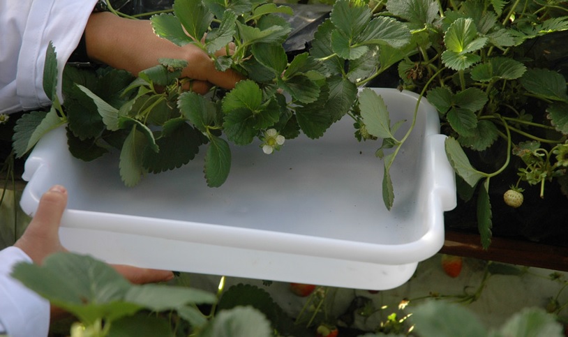
To collect thrips in plants, beating the vegetation over a plastic tray (preferably white) is one of the most used techniques. After falling in the tray, thrips can be collected carefully with a fine brush, wet with 60% alcohol, and transferred immediately into an Eppendorf tube. To collect thrips from flowers with a narrow opening, dense inflorescences (e. g. Asteraceae) or galls it is recommended the inspection of these structures in laboratory with the use of a stereomicroscope. Beating or sweeping nets may also be used for collecting thrips in plants, however the efficiency is more limited and screening the material is slower; moreover, thrips taken with sweepnets are frequently damaged.
To collect and study fungivorous thrips it is needed to inspect tree bark, dead twigs, leaf litter and fallen tree trunks. For dry twigs and dead leaves still hanging from the plant it is reasonable to adopt the same technique of beating over a plastic tray described above. It is possible to capture thrips inhabiting the leaf litter with Pitfall traps, or transferring dry leaves samples to a Berlese funnel for at least 24 hours. A good method of collecting thrips from tree bark and fallen branches is to spray the surface with a domestic insectide. After a few minutes, insects can be collected as they fall onto a sheet laid below.
Thrips have a fragile body and need to be manipulated with care, to avoid losing structures important for identification. The alcohol concentration is also an important variable for thrips preservation. Usually thrips are conserved in 60% alcohol, but it is possible to prepare good microscopic slides with individuals retained in 70-95% alcohol. However, specimens conserved in concentrations above 80% are more susceptible to being damaged by manipulation. In such cases, we reccomend rehydratation of the specimens in destilled water for at least 6h before maceration.
The collection data with date, local, plant/environment may be written in a tracing paper strip and placed inside the tube, together with the specimens.
Thrips may also be collected into AGA (10 parts of 60% ethyl alcohol, one part of glycerine and one part of glacial acetic acid), which helps to preserve the malleability of specimens. It is important to observe that the usage of AGA is not indicated for samples that will be used in molecular analysis.
Preparing thrips for study
Thrips can be prepared for study in different ways, but for most studies they must be mounted onto microscopic slides. Two techniques are commonly used, depending on the objectives of the activity: the semi-permanent slides, which use a mounting media based on water, and the permanent slides, which frequently use Canada Balsam.
Preparing semi-permanent slides is fast and efficient for routine identification with the need of preserving the material for longer periods. The mounting media named Hoyer’s is most frequently used by specialists.
Preparation of Hoyer’s mounting media:
Distilled water....................40 ml
Gum Arabic (in crystals)....30 g
Chloral hydrate..................200 g
Glycerine.............................20 ml
These substances must be mixed in this order and in room temperature, and afterwards filtered in cotton or gauze. After preparing this mixture, it is recommended to wait at least 24h to use it, so the air bubbles can leave the liquid.
To start the mounting process specimens should be macerated to remove internal contents. This and the following procedures must be performed using a stereomicroscope.
Below follows a preparation protocol for semi-permanent slides used by the authors:
1-Place the thrips in a small tube or petri dish containing KOH 5-8%. Thrips must remain in the liquid for at least 3 hours, sometimes longer if a specimen is dark or even black (some thrips must remain for over 24 hours). During this period pierce the abdomen of the thrips in the ventral surface, using a micro pin, preferably between the posterior coxae, and gently rub the abdomen to remove the internal contents.
2-Put the thrips in distilled water for 10 minutes to remove the KOH.
3-Transfer the specimens to 60% alcohol for about 24 hours.
4-Open and straighten the legs, antennae and wings of thrips.
5-Place a droplet of Hoyer’s over a microscope cover slip (15x15 mm or smaller).
6-Transfer the thrips to the Hoyer’s droplet with the help of a micro pin, positioning the specimen in the centre of the cover slip with the ventral region facing upwards.
7-Put a droplet of Hoyer’s in the middle of a microscope slide and touch it to the Hoyer’s on the cover slip (liquid with liquid), without releasing the slide over the slip. After the liquids spread, turn the slide upside down quickly, such that the cover slip it is uppermost.
8-Immediately place the slide over a previously heated surface at 50°C, and let the slide rest there for some hours before observing it with a microscope.
Remember that Hoyer’s needs some months to dry, so slides must be kept horizontal, not vertical. It is recommended to put the slides inside an incubator at 50°C to accelerate drying the mounting media. The use of a varnish or enamel to seal the edges of the coverslip is recommended to increase durability.
Now you may identify your material clicking here.
For taxonomic studies of thrips, it is recommended to prepare permanent slides, mounted in Canada Balsam. Slides prepared with this mounting media have unknown durability, but slides prepared over 100 years ago are still well preserved.
Mounting in Canada Balsam requires a dehydration process for the specimens, or else the material may be damaged, or bubbles may be formed, impairing observation.
Similar to the preparation in Hoyer’s, thrips must be macerated in KOH 5% following the steps 1-4 from the protocol described above. We recommend the following protocol for preparing permanent slides:
1-Transfer the macerated thrips to 60% alcohol for at least 30 min.
2-Transfer the macerated thrips to 70% alcohol for 1 hour.
3-Transfer the thrips to 80% alcohol for 20 minutes.
4-Transfer the thrips to 90-95% alcohol for 10 minutes.
5- Transfer the thrips to 100% alcohol for 10 minutes.
6-Transfer the thrips to clove oil (Eugenol) for at least 1 hour. Do not use clove oil diluted in water, largely used as an aromatic essence.
7- Place a droplet of Canada Balsam over a microscopic cover slip (15x15 mm or smaller). The Balsam used for mounting must not be thick, which could damage the specimen. We recommend diluting it using xylene.
8-Transfer one (or more) thrips to the droplet with the help of a micro pin, positioning the specimen in the centre of the cover slip with the ventral region facing upwards.
9-Put a droplet of Canada Balsam in the middle of a microscope slide and touch it to the cover slip (liquid with liquid), without releasing the slide over the slip. After the liquids spread, turn the slide upside down quickly, such that the cover slip it is uppermost.
10-Immediately place the slide over a previously heated surface at 50°C, and let the slide rest there for some hours before observing it under a microscope.
Important: the amount of mounting media in both types of slide preparation must be sufficient to support the cover slip pressure without distorting the specimen.
The slides need some months to dry, thus they must be kept horizontal not in a vertical position. It is recommended to put the slides inside an incubator at 50°C to accelerate drying the mounting media.
The slides must be properly labelled for taxonomic and storage purposes, and the use of adhesive labels facilitates this procedure. Usually, the label at the left side of the slide has the identification data, while the label on the right side has the collection data.
Attendance
Brasil, Rio Grande do Sul, Rio Grande
Campus Carreiros: Av. Itália km 8
Bairro Carreiros
Thysanoptera © Todos Direitos Reservados 2026